Labeled Diagram Of An - Well Labeled Diagram Of The Grapes Components Download Scientific Diagram / Label fractions on number line.. The diagram below shows the structure and functions of the human digestive system. The system breaks down food, extracts nutrients from it, and converts them into energy. These bones are arranged into two major divisions: Label the drawing of the crayfish. We hope this picture labelled diagram of the muscles in the human body can help you study and research.
In a plant cell, the cell wall is made up of cellulose, hemicellulose, and proteins while in a fungal cell, it is composed of chitin. Let learn the different parts of the human digestive system. As shown in the diagram on the next page, its body is divided into two main parts, the cephalothorax and the abdomen. Anatomynote.com found labelled diagram of the muscles in the human body from plenty of anatomical pictures on the internet. Labeled diagram of the human lungs.
Overview Of Keys Labeled Diagram Of The Keyboard Transparent Png Download 1311628 Vippng from www.vippng.com As shown in the diagram on the next page, its body is divided into two main parts, the cephalothorax and the abdomen. The system breaks down food, extracts nutrients from it, and converts them into energy. Label the drawing of the crayfish. In a typical animal cell, mitosis can be divided into stages: Mitosis is a process of cell division which results in the production of two daughter cells from a single parent cell. We hope this picture labelled diagram of the muscles in the human body can help you study and research. In a plant cell, the cell wall is made up of cellulose, hemicellulose, and proteins while in a fungal cell, it is composed of chitin. The daughter cells are identical to one another and to the original parent cell.
Anatomynote.com found labelled diagram of the muscles in the human body from plenty of anatomical pictures on the internet.
Let learn the different parts of the human digestive system. The axial skeleton and the appendicular skeleton. Diagram of human heart and blood circulation in it heart is a vital organ that you cannot live without. You should now be able to label all the main anatomical features. They are found in the brain, spinal cord and the peripheral nerves. The heart, one of the most significant organs in the human body, is nothing but a muscular pump which pumps blood throughout the body. We think this is the most useful. The function of heart is quite complex, but you can understand things better through the heart diagram labeled below. Labelled diagram drag and drop the pins to their correct place on the image. It is the beginning of the digestive tract and the process of digestion begins from the mouth, where teeth help by breaking and grinding the food. The heart, though small in size, performs highly significant functions that sustains human life. The structure of a neuron varies with their shape and size and it mainly depends upon their. To understand one of the most complex joints of our body i.e.
They are found in the brain, spinal cord and the peripheral nerves. Middle lamina contains polysaccharides that provide adhesion and allows binding of the cells to one. An atom is the basic unit of matter. (physical cues of anger) by zazaef. Like all crustaceans, a crayfish has a fairly hard exoskeleton that covers its body.
Femur Bone Anatomy Labeled Diagram Quiz Color Coded Parts Skeletal System Lower Extremity Ezmed from images.squarespace-cdn.com Label fractions on number line. They are found in the brain, spinal cord and the peripheral nerves. Anatomynote.com found labelled diagram of the muscles in the human body from plenty of anatomical pictures on the internet. This diagram depicts labeled diagram of digestive system.human anatomy diagrams show internal organs, cells, systems, conditions, symptoms and sickness information and/or tips for healthy living. The function of heart is quite complex, but you can understand things better through the heart diagram labeled below. The human digestive system is the means by which tissues and organs receive nutrients to function. These bones are arranged into two major divisions: The heart, one of the most significant organs in the human body, is nothing but a muscular pump which pumps blood throughout the body.
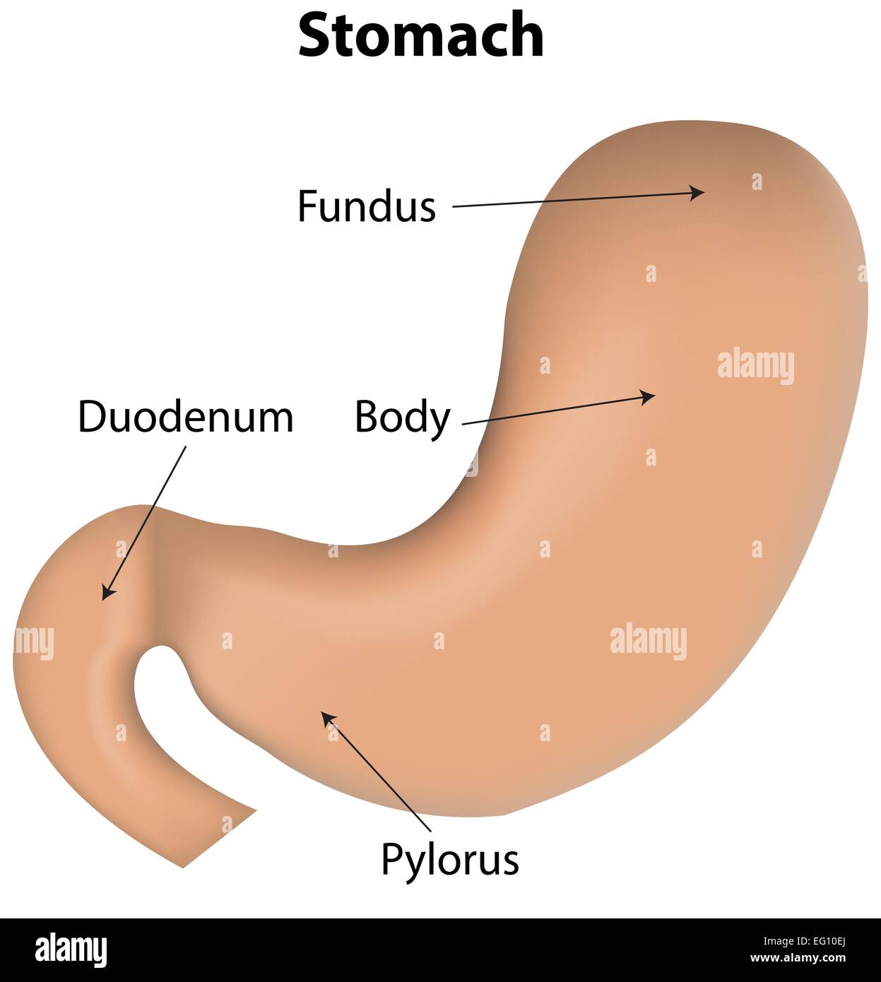
It is the beginning of the digestive tract and the process of digestion begins from the mouth, where teeth help by breaking and grinding the food.
The human heart and its functions are truly fascinating. In addition, they also play an important role in maintaining the water balance of our body. Below is a blank diagram, followed by the labeled diagram with the answers. In a plant cell, the cell wall is made up of cellulose, hemicellulose, and proteins while in a fungal cell, it is composed of chitin. We think this is the most useful. The function of heart is quite complex, but you can understand things better through the heart diagram labeled below. (physical cues of anger) by zazaef. We hope this picture labelled diagram of the muscles in the human body can help you study and research. Mitosis is a process of cell division which results in the production of two daughter cells from a single parent cell. It is the beginning of the digestive tract and the process of digestion begins from the mouth, where teeth help by breaking and grinding the food. Illustration of the process by which somatic cells multiply and divide. Let learn the different parts of the human digestive system. To understand one of the most complex joints of our body i.e.
Illustration of the process by which somatic cells multiply and divide. A labeled diagram of the human heart you really need to see. The axial skeleton and the appendicular skeleton. Labeled diagram of the human kidney. The following article provides you with diagrams that will help you understand the structure of an atom better.
Stomach Labeled Diagram Stock Vector Image Art Alamy from c8.alamy.com The daughter cells are identical to one another and to the original parent cell. The function of heart is quite complex, but you can understand things better through the heart diagram labeled below. Label fractions on number line. The heart, one of the most significant organs in the human body, is nothing but a muscular pump which pumps blood throughout the body. Middle lamina contains polysaccharides that provide adhesion and allows binding of the cells to one. Illustration of the process by which somatic cells multiply and divide. We think this is the most useful. Labeled diagram of the human kidney.
The heart, one of the most significant organs in the human body, is nothing but a muscular pump which pumps blood throughout the body.
They are found in the brain, spinal cord and the peripheral nerves. Diagram of human heart and blood circulation in it heart is a vital organ that you cannot live without. The following article provides you with diagrams that will help you understand the structure of an atom better. The function of heart is quite complex, but you can understand things better through the heart diagram labeled below. A labeled diagram of the knee with an insight into its working. This diagram depicts labeled diagram of digestive system.human anatomy diagrams show internal organs, cells, systems, conditions, symptoms and sickness information and/or tips for healthy living. Who let the hulk out? You should now be able to label all the main anatomical features. Middle lamina contains polysaccharides that provide adhesion and allows binding of the cells to one. Label fractions on number line. In addition, they also play an important role in maintaining the water balance of our body. Labelled diagram drag and drop the pins to their correct place on the image. In a plant cell, the cell wall is made up of cellulose, hemicellulose, and proteins while in a fungal cell, it is composed of chitin.


0 Komentar